
Sometimes scientists start out researching one subject, but along the way, they come across something else even more interesting.
This is what happened to University of Georgia Skidaway Institute of Oceanography researcher Adam Greer in the summer of 2016 when Greer was a post-doctoral associate at the University of Southern Mississippi.
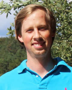
Adam Greer
That fortuitous event resulted in a paper recently published in the journal Limnology and Oceanography with Greer as the lead author.
Greer and his fellow researchers were on a cruise in the northern Gulf of Mexico to study the effects of river input on biological processes. They came across a natural phenomenon called a thin layer. These are oceanographic features found all over the world where biomass collects into a narrow portion of the water column–less than five meters thick vertically–and can extend for several kilometers horizontally. They tend to occur in stratified shelf systems.
“Surprisingly, there are few published studies on thin layers in the northern Gulf of Mexico, which is heavily influenced by rivers and highly stratified during the summer,” Greer said. “Thin layers are important because they are trophic hot spots, where life tends to congregate, and predators and prey interact.”
However, Greer said, thin layers are very difficult to analyze because they occur within a restricted portion of the water column, and most conventional ocean sampling equipment will not detect their influence on different organisms.
Greer and his colleagues were better equipped than most to study the thin layer. Rather than laying out a grid and taking a series of water samples, they were equipped with an In Situ Ichthyoplankton Imaging System (ISIIS).
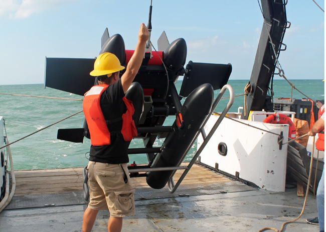
Launching the In Situ Ichthyoplankton Imaging System from the deck of a research vessel.
This imaging system was towed behind their research vessel and undulated through the water column, producing a live feed of plankton images and oceanographic data. By studying the video, they were able to map the distributions of many different types of organisms in great detail. The thin layer was composed of large chains of phytoplankton called diatoms and gelatinous zooplankton called doliolids.
“Although we expected many different organisms to aggregate within the layer, this was not the case,” Greer said. “The only organisms that were concentrated within the layer were gelatinous organisms called doliolids. Other organisms that we expected to see, such as copepods, chaetognaths and shrimp, tended to congregate near the surface just south of the thin layer.”
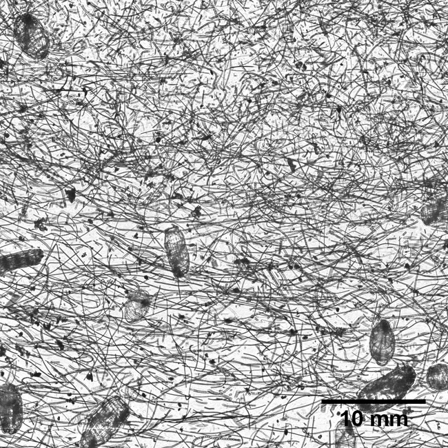
An image from the In Situ Ichthyoplankton Imaging System passing through the thin layer. The long, slender filaments are chains of diatoms. The larger, oval plankton are doliolids.
The researchers determined that the area south of the thin layer was influenced by a surface convergence – two water masses colliding and pushing water downward at a slow rate. They believe that many organisms with active swimming ability, such as shrimps and copepods, could stay within the surface convergence, while more passive swimmers, such as doliolids would follow the trajectory of the thin layer and diatoms.
Greer and his colleagues discovered several other characteristics of the thin layer they had not anticipated. There was a higher concentration of live phytoplankton than expected. As a result, the thin layer also had a high concentration of dissolved oxygen due to the photosynthetic activity. The zooplankton were also aggregated into distinct microhabitats with different oceanographic properties — such as temperature, salinity and light. The microhabitats also contained different types and abundances of food.
“For a lot of these organisms, if you took the average abundance of food it wouldn’t be enough to survive,” Greer said. “So whatever mechanisms there are to create higher abundances of food, they are potentially really important for a number of different organisms.”
The other members of the research team were Adam Boyette, Valerie Cruz, Kemal Cambazoglu, Luciano Chiaverano and Jerry Wiggert, all from the University of Southern Mississippi; Brian Dzwonkowski and Steven Dykstra, from the University of South Alabama; and Christian Briseño‐Avena and Bob Cowen, from Oregon State University.
The paper can be viewed HERE.
An image from the In Situ Ichthyoplankton Imaging System passing through the thin layer. The long, slender filaments are chains of diatoms. The larger, oval plankton are doliolids